


pCD2-hTL1A-Tg
品系全名
C57BL/6JSmo-Tgtn(pCD2-hTL1A)1Smoc
目录号
NM-TG-250279
品系状态
精子冻存
品系描述
验证数据

Fig.1 Detection of human TL1A expression in serum by ELISA (n=3, 6 weeks old).
Abbr. WT, wild type; N.D. not detected.
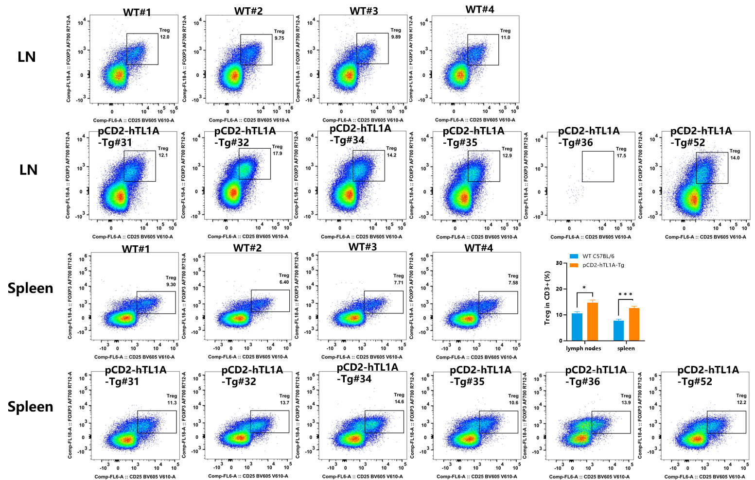
Fig.2 Detection of Treg expression on CD3+T cells in MLN and spleen of WT (C57BL/6) mice and pCD2-hTL1A-Tg mice by FACS.
In CD3+ T cells, compared with wild-type mice, the number of Treg cells in the spleen and MLN of pCD2-hTL1A-Tg mice was increased (n=4-6, 17 weeks old).
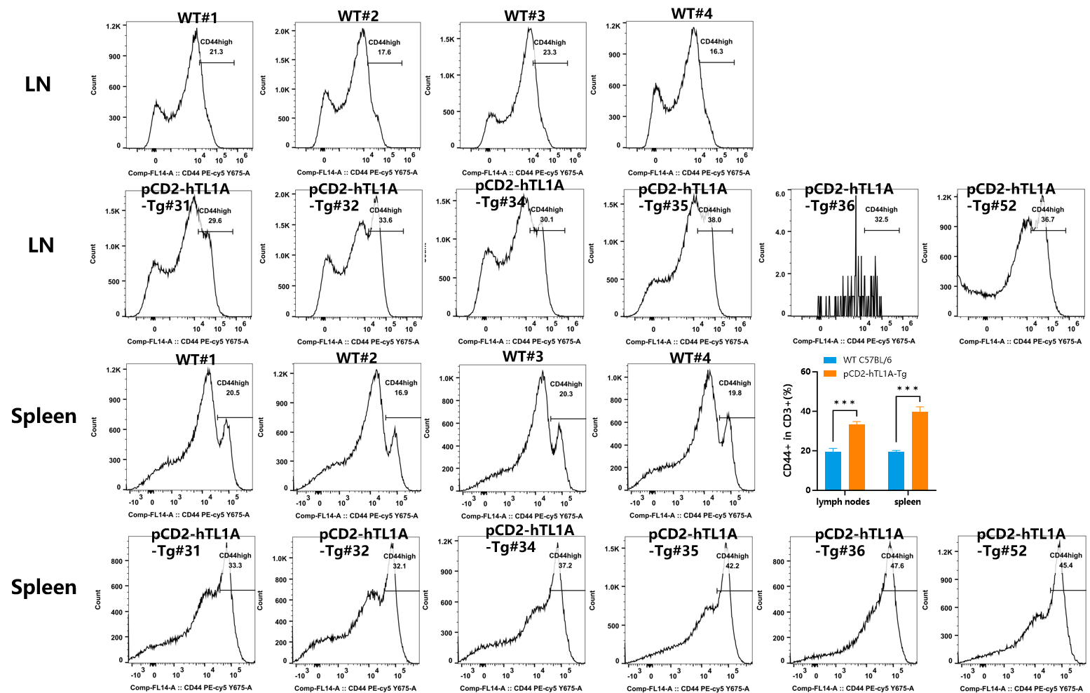
Fig.3 Detection of CD44 expression on CD3+T cells in MLN and spleen of WT (C57BL/6) mice and pCD2-hTL1A-Tg mice by FACS.
In CD3+ T cells, compared with wild-type mice, the number of CD44+ cells in the spleen and MLN of pCD2-hTL1A-Tg mice was increased (n=4-6, 17 weeks old).
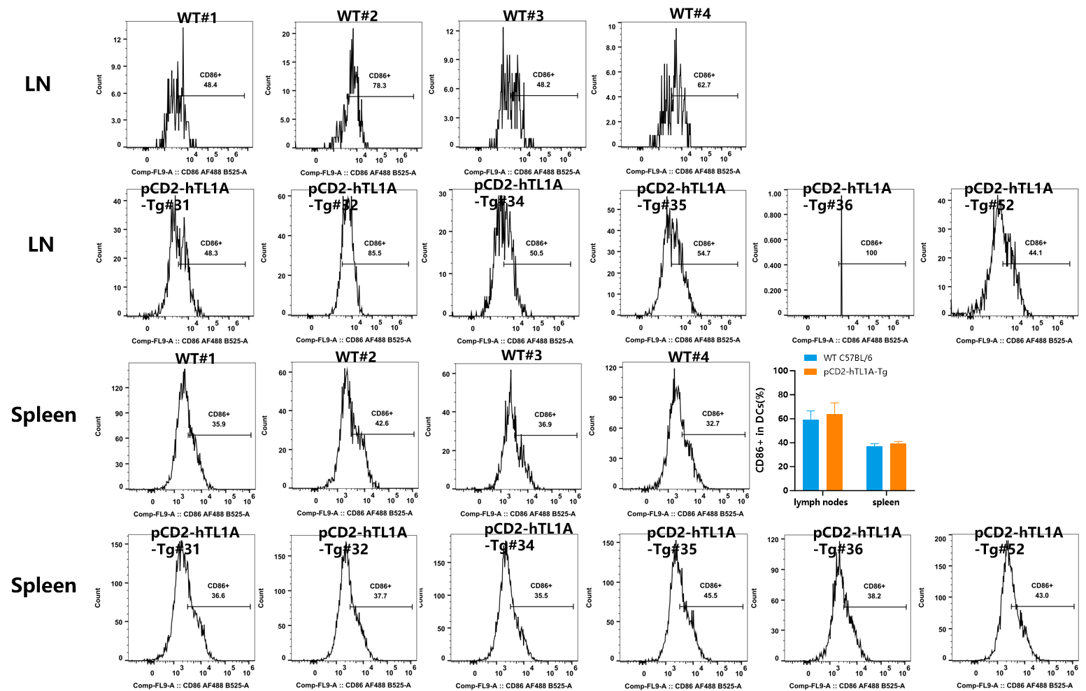
Fig.4 Detection of CD86 expression on CD11c+DCs in MLN and spleen of WT (C57BL/6) mice and pCD2-hTL1A-Tg mice by FACS.
In DCs cells, the number of CD86+ cells in the spleen and MLN of pCD2-hTL1A-Tg mice was similar to that of wild-type mice (n=4-6, 17 weeks old).
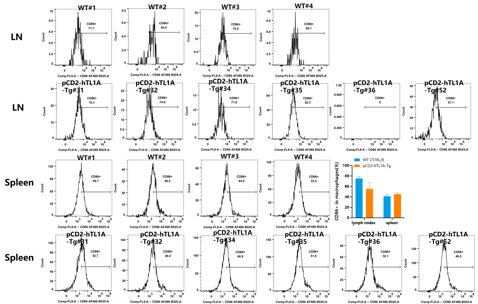
Fig.5 Detection of CD44 expression on macrophages in MLN and spleen of WT (C57BL/6) mice and pCD2-hTL1A-Tg mice by FACS(n=4-6, 17 weeks old).
In macrophages, the number of CD86+ cells in the spleen and MLN of pCD2-hTL1A-Tg mice was similar to that of wild-type mice.


Fig.6 Detection of IFN-γ expression on CD4+T cells in MLN and spleen of WT (C57BL/6) mice and pCD2-hTL1A-Tg mice by FACS.
In CD4+ T cells, compared with wild-type mice, the number of IFNγ+ cells in the spleen and MLN of pCD2-hTL1A-Tg mice was significantly increased (n=4-6, 17 weeks old).

Fig.7 Detection of IL-17A+ expression on CD4+T cells in MLN and spleen of WT (C57BL/6) mice and pCD2-hTL1A-Tg mice by FACS.
In CD4+ T cells, compared with wild-type mice, the number of IL-17A+ cells in the spleen and MLN of pCD2-hTL1A-Tg mice was significantly increased (n=4-6, 17 weeks old).

Fig.8 Detection of IL-4+ expression on CD4+T cells in MLN and spleen of WT (C57BL/6) mice and pCD2-hTL1A-Tg mice by FACS.
No IL-4+ cells were observed in the spleen and MLN of pCD2-hTL1A-Tg mice and wild-type mice (n=4-6, 17 weeks old).

Fig. 8 Phenotype of WT (C57BL/6) mice and pCD2-hTL1A-Tg mice (n=2-3, 17 weeks old). (A) Gross photograph; (B) Colon photo; (C) Body weight and intestine index.

Fig. 9 H&E staining of the small intestine and colon in pCD2-hTL1A-Tg mice (n=2-3, 17 weeks old).
你也可能感兴趣
Tamoxifen诱导Cre-ERT2小鼠 使用指南
Cre-ERT2在无Tamoxifen诱导的情况下,在细胞质内处于无活性状态;当Tamoxifen诱导后,Tamoxifen的代谢产物4-OHT(雌激素类似物)与ERT结合,可使Cre-ERT2进核发挥Cre重组酶活性。
查看Cre-lox系统介绍及使用汇总
你一定听说过Cre-lox重组系统,无论你是否直接进行过基因操作。由于Cre-lox系统具有操作简单、重组率高的优点,如今已经成为体内外遗传操作的强有力工具。利用Cre-lox系统,可以在特定细胞、组织或整个生物体,甚至在特定时间点敲除或表达某个基因,实现对特定基因的时空特异性操作,这对基因功能的研究和人类疾病动物模型的建立都具有深刻影响。
查看


